Examples
Complete studies, each run end to end on a GPU: the code, the prose and every figure, rendered in place. Each page links to the notebook's source, which opens in Pluto.
Getting started
The API end to end, on one clinical scanner.
Phantoms & anatomy
Digital anatomy, custom materials, and mapping ground truth onto the reconstruction.
XCAT Anatomy + Custom Materials
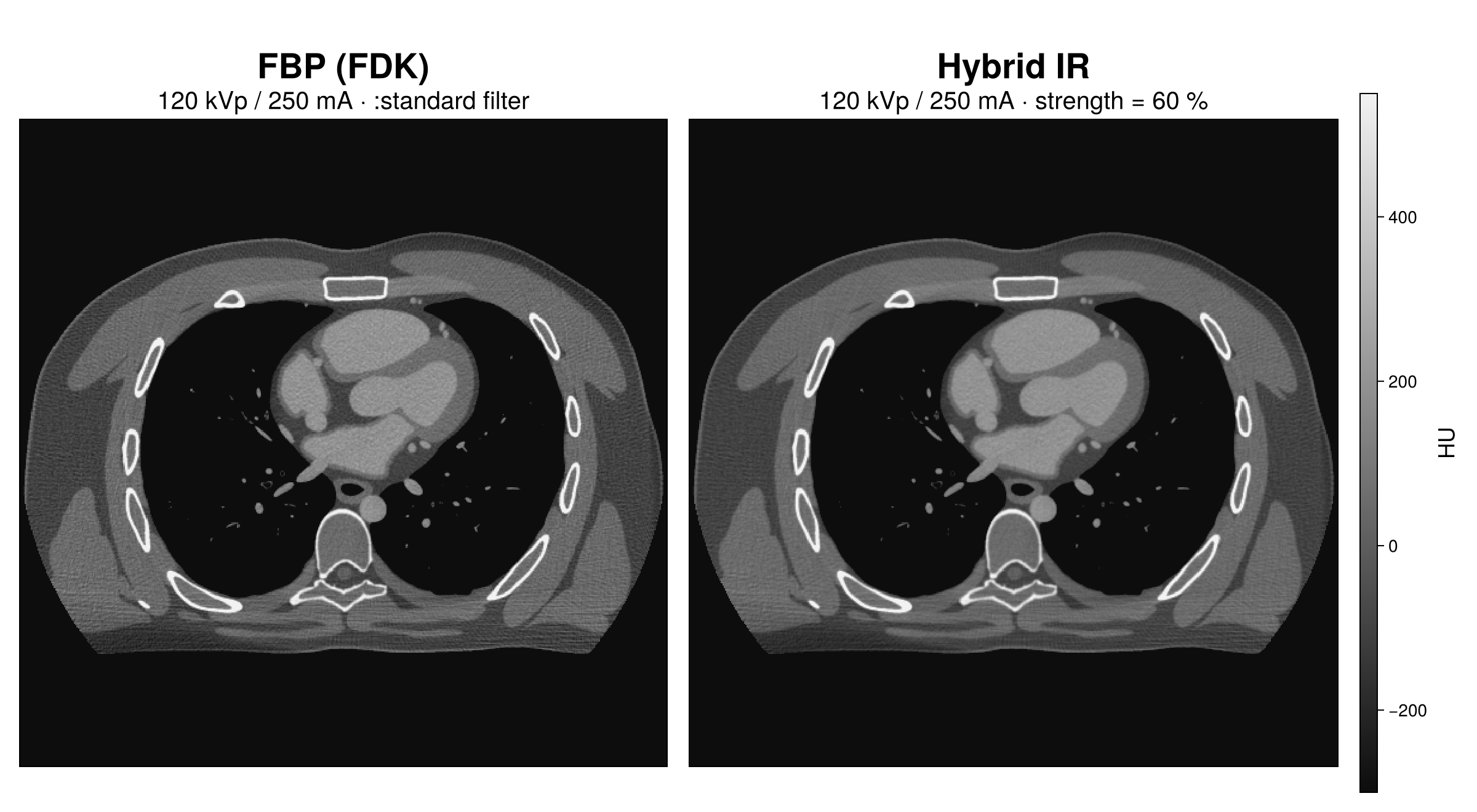
An XCAT adult-male chest with an XrayAttenuation material for every organ, including an iodinated blood mixture defined inline, scanned on the Apex Elite and reconstructed with FBP and hybrid IR at strength 60 after the water beam-hardening correction.
XCAT Grids and the Affine Round-Trip
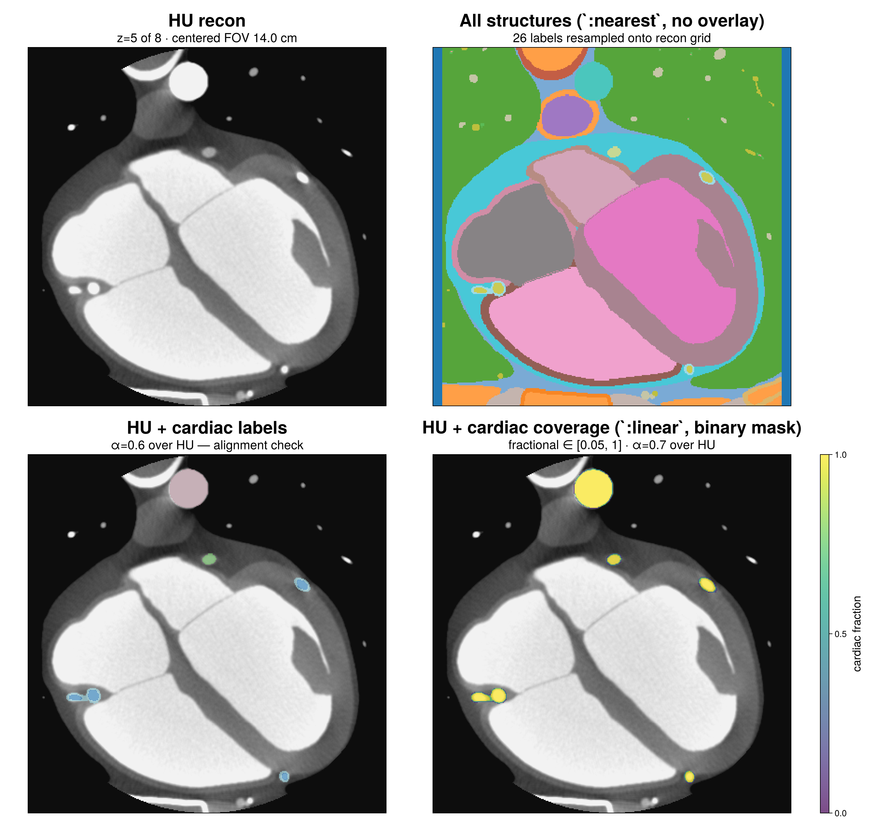
An XCAT 3.0 male chest export cropped to the heart and scanned axially and helically; phantom_to_world_affine, recon_to_world_affine and resample_to_recon register the ground-truth labels onto both reconstructions, with organ ROI statistics and an exactness audit.
Spectral & VMI
Dual-kVp and photon-counting acquisitions through BS.vmi_pipeline, checked rod by rod against theory.
Dual-kVp Switching VMI
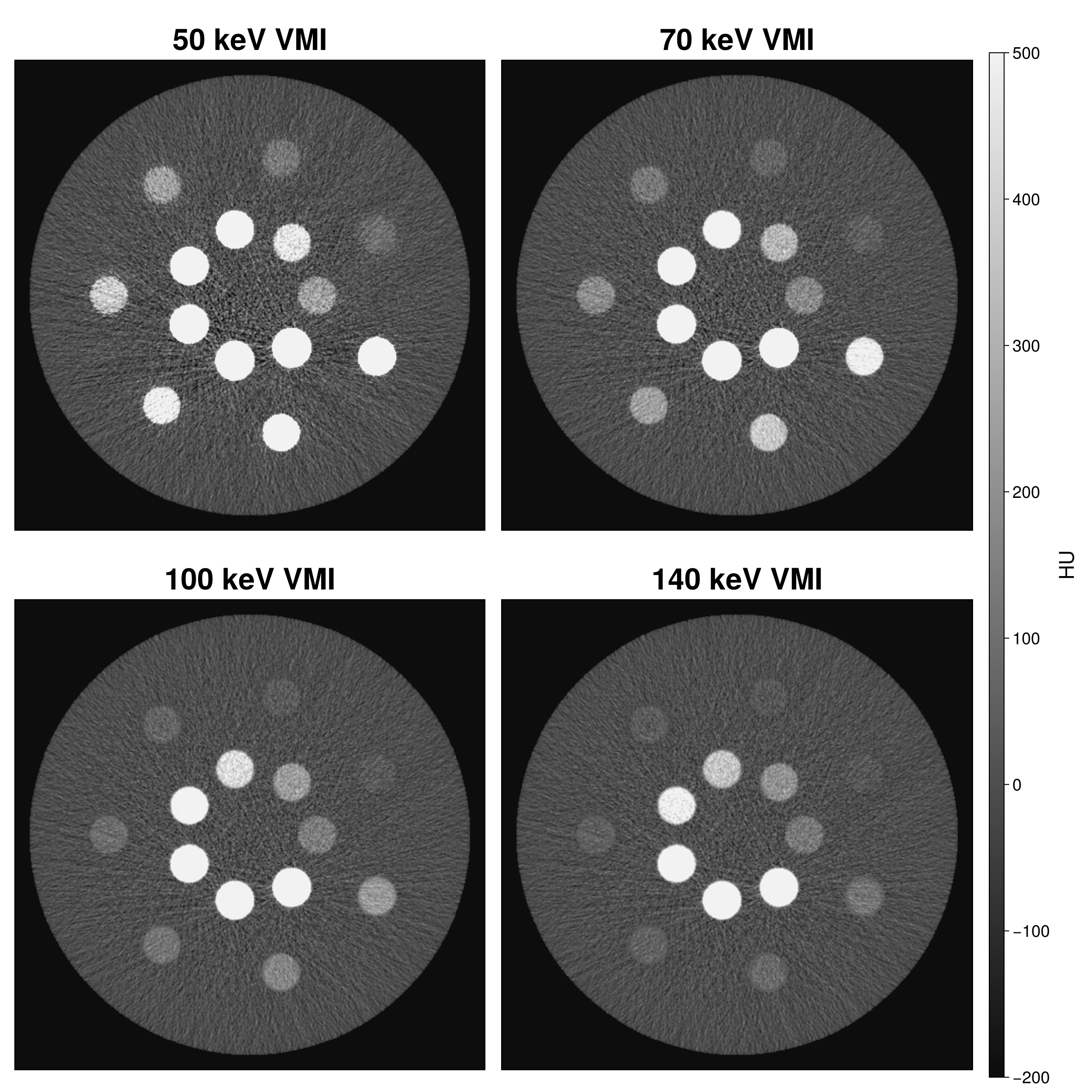
GE Apex Elite rapid kVp switching (80/140 kVp) on Gammex 472 through BS.vmi_pipeline: n-channel profile-likelihood decomposition, SpectralHYPR in the projection and image domains, ACNR, and VMIs at 50, 70, 100 and 140 keV checked rod by rod against theory.
Photon-Counting VMI
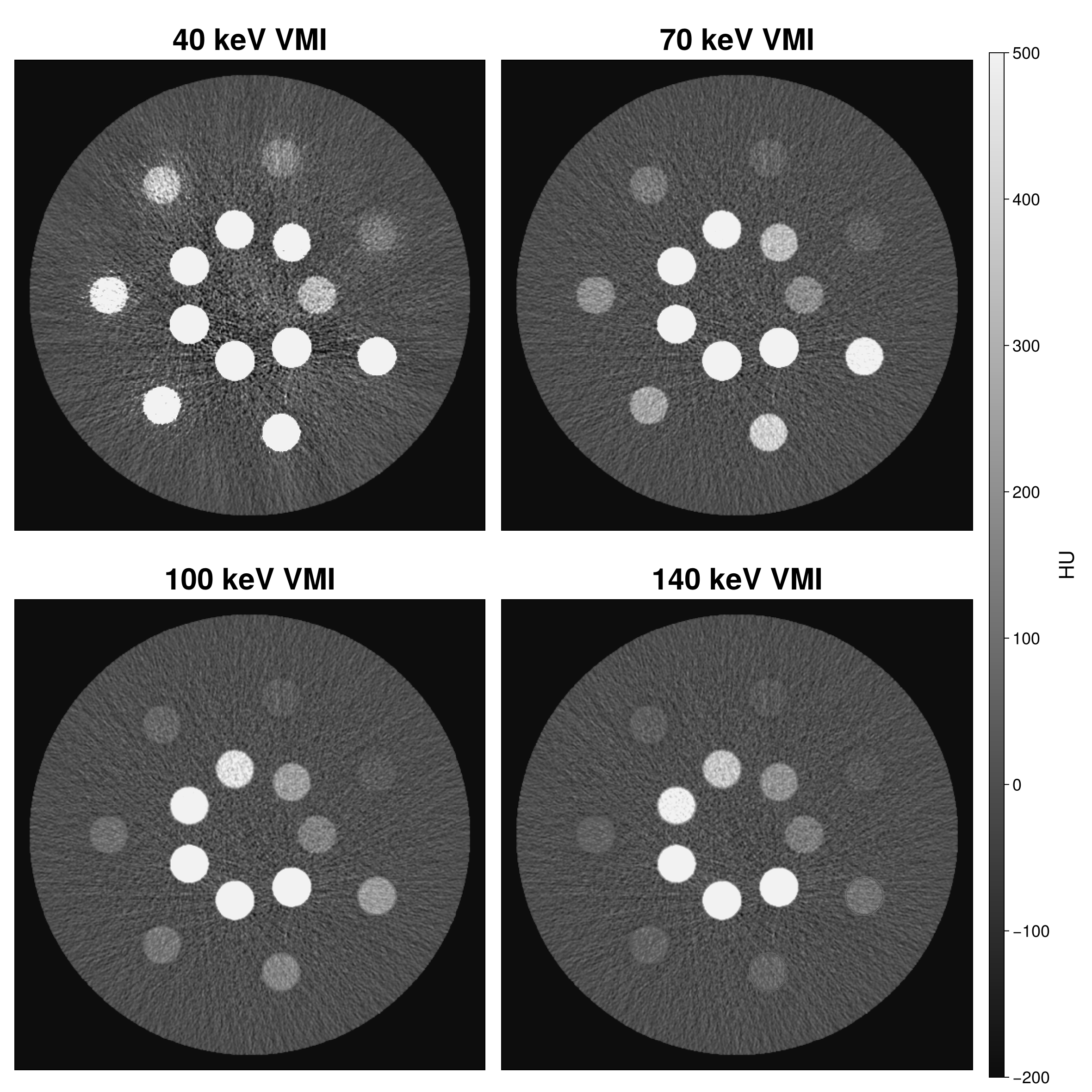
A Siemens NAEOTOM Alpha model at 140 kVp with four counting bins on Gammex 472: the per-ray spectral_basis(ws; I0) of the simulation, one BS.vmi_pipeline call, and VMIs at 40, 70, 100 and 140 keV verified rod by rod against theory.
QRM Thorax: Pure-Material Dual-kVp VMI
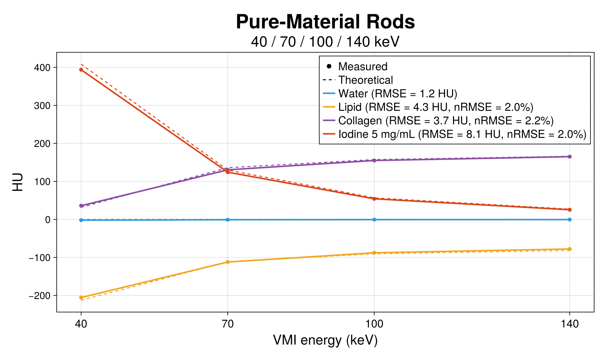
A body-sized QRM thorax phantom, built analytically in the notebook, with four pure-material rods (water, lipid, collagen, iodine), scanned by Apex Elite rapid kVp switching at clinical resolution; BS.vmi_pipeline and a per-rod measured-versus-theoretical regression.
QRM Thorax: Pure-Material Photon-Counting VMI
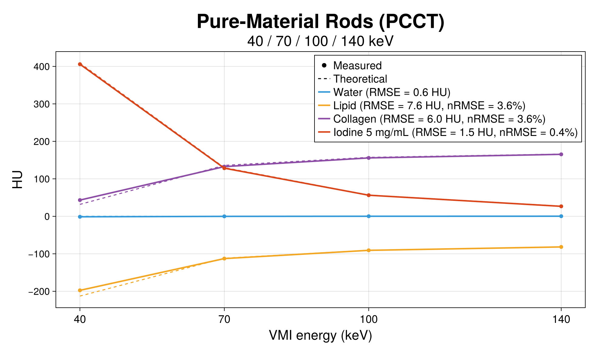
The photon-counting counterpart of notebook 07: the same analytic thorax phantom and rods, one 140 kVp NAEOTOM Alpha acquisition with pile-up and scatter correction in the detector model, BS.vmi_pipeline, and the same per-rod regression.
Scanner models
Dual-source Siemens systems with their own Monte Carlo detector tables, read out as mixed images and VMIs.
Siemens SOMATOM Force: Dual-Source VMI
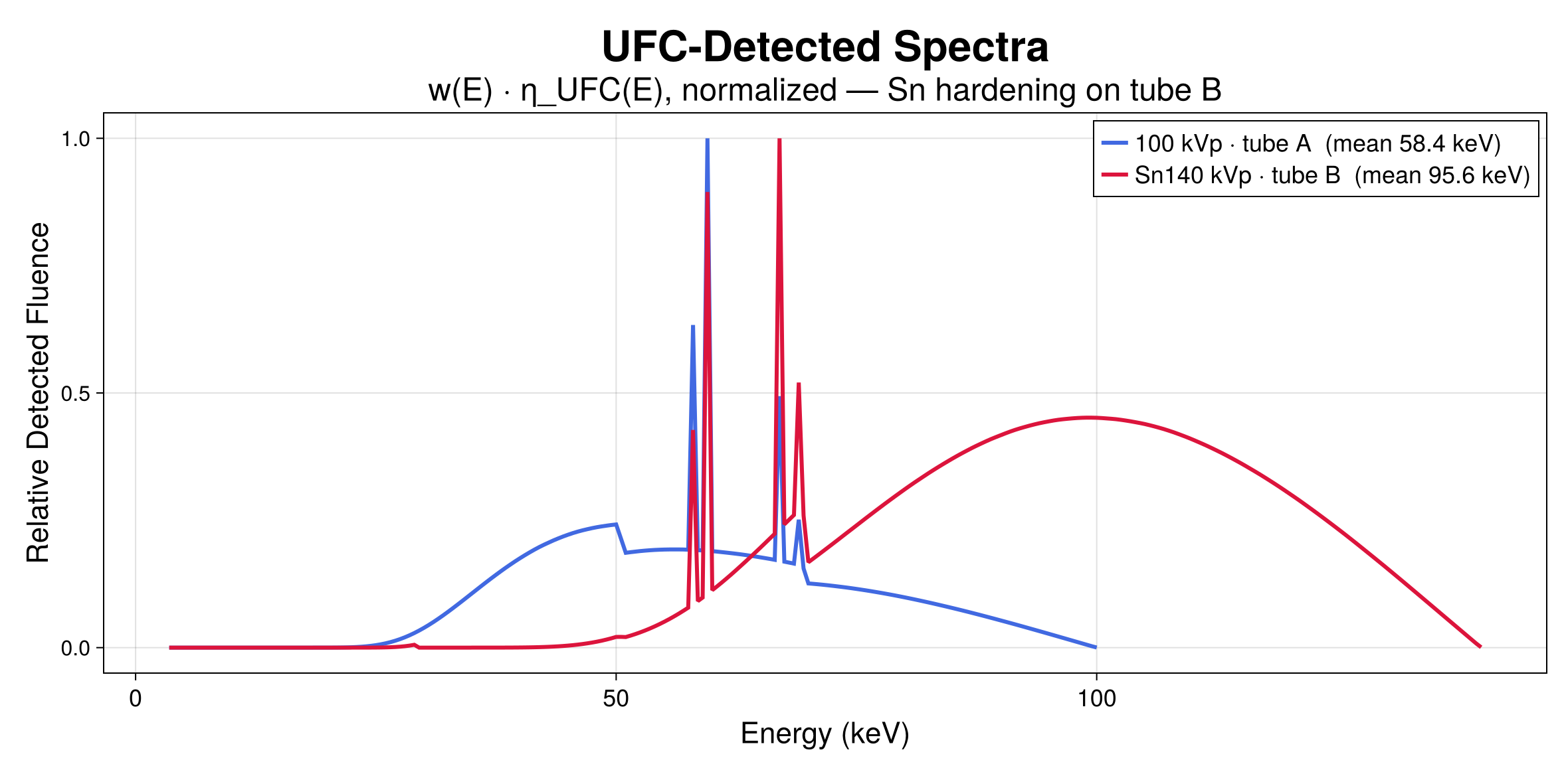
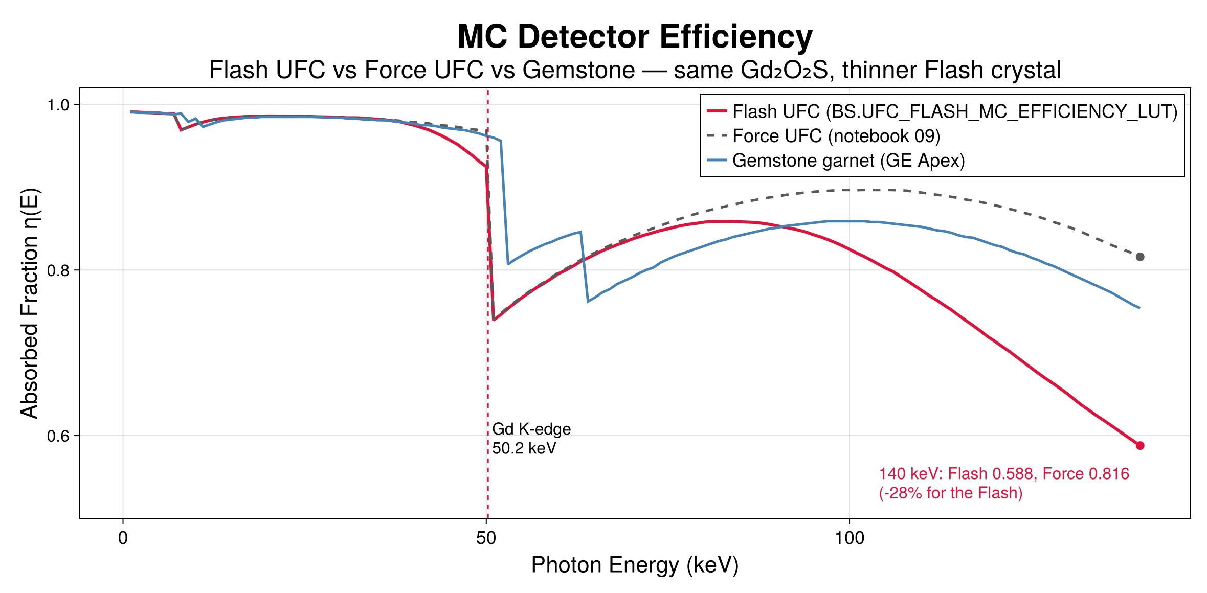
The third-generation dual-source Force with its UFC Gd₂O₂S Monte Carlo efficiency table. One 100/Sn140 kV acquisition read out two ways: per-tube reconstructions with a Siemens-style mixed image, and BS.vmi_pipeline VMIs checked per rod.
Siemens SOMATOM Definition Flash
The second-generation dual-source Flash with its own, thinner UFC crystal table. Regular dual-power scanning at 120 kV (water accuracy and the √2 noise gain of two tubes) and the clinical 80/Sn140 kV pair to a mixed image and BS.vmi_pipeline VMIs, closed by a 21-check verification gate.
Acquisition, artifacts & validation
Helical coverage, metal artifacts, and a head-to-head with CatSim.
Helical Scanning
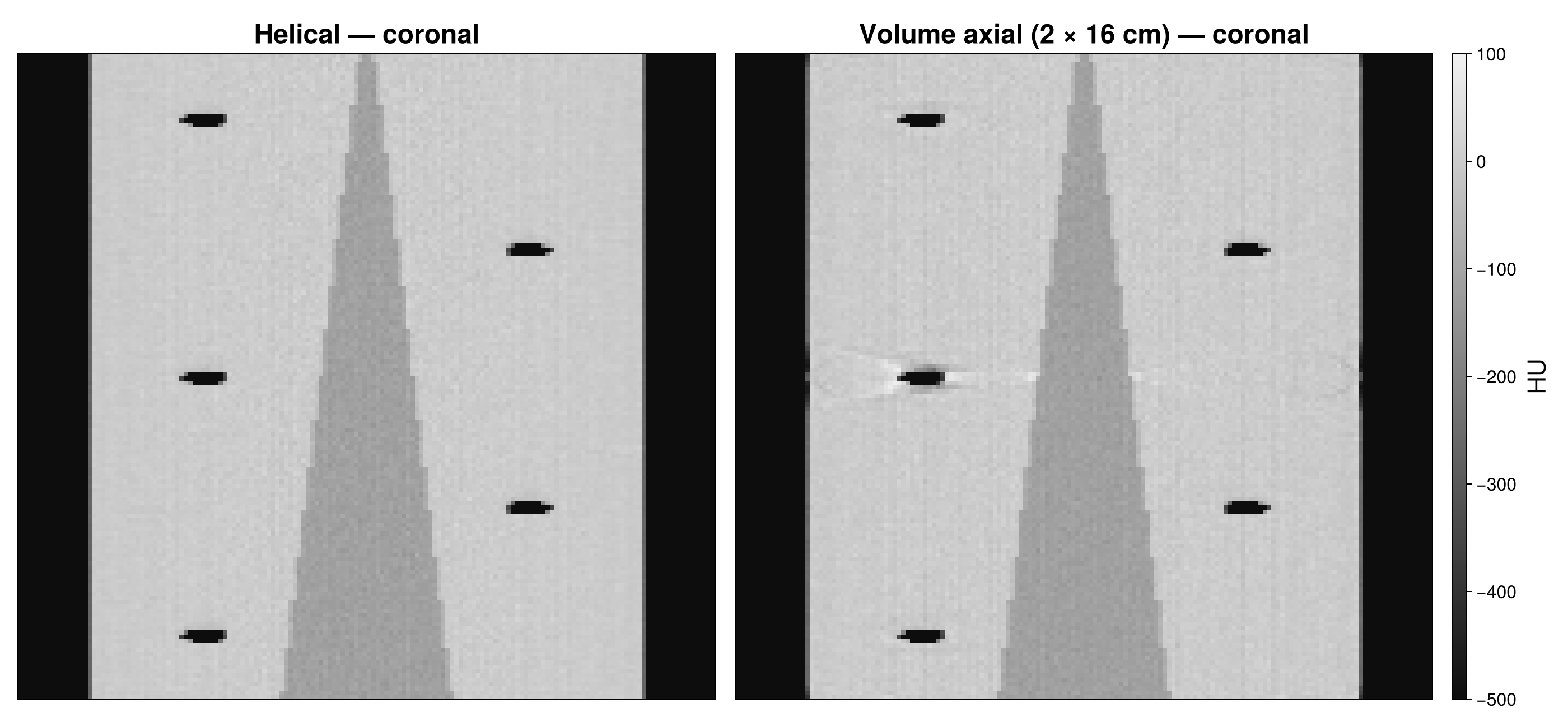
One keyword, pitch, turns an axial protocol into a spiral. A 30 cm slab from a 20 mm helix at pitch 1.0 against three volume-axial stations (16 cm beam, 10 cm apart) at matched beam-width × current: rebinned WFBP, CTDIvol and DLP of each, coronal reformats and z-profiles.
Titanium Implant Artifacts
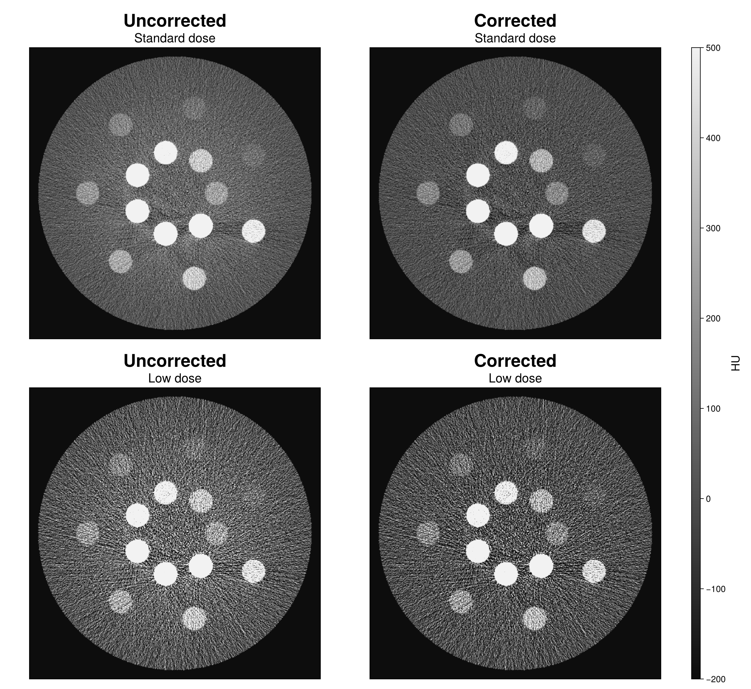
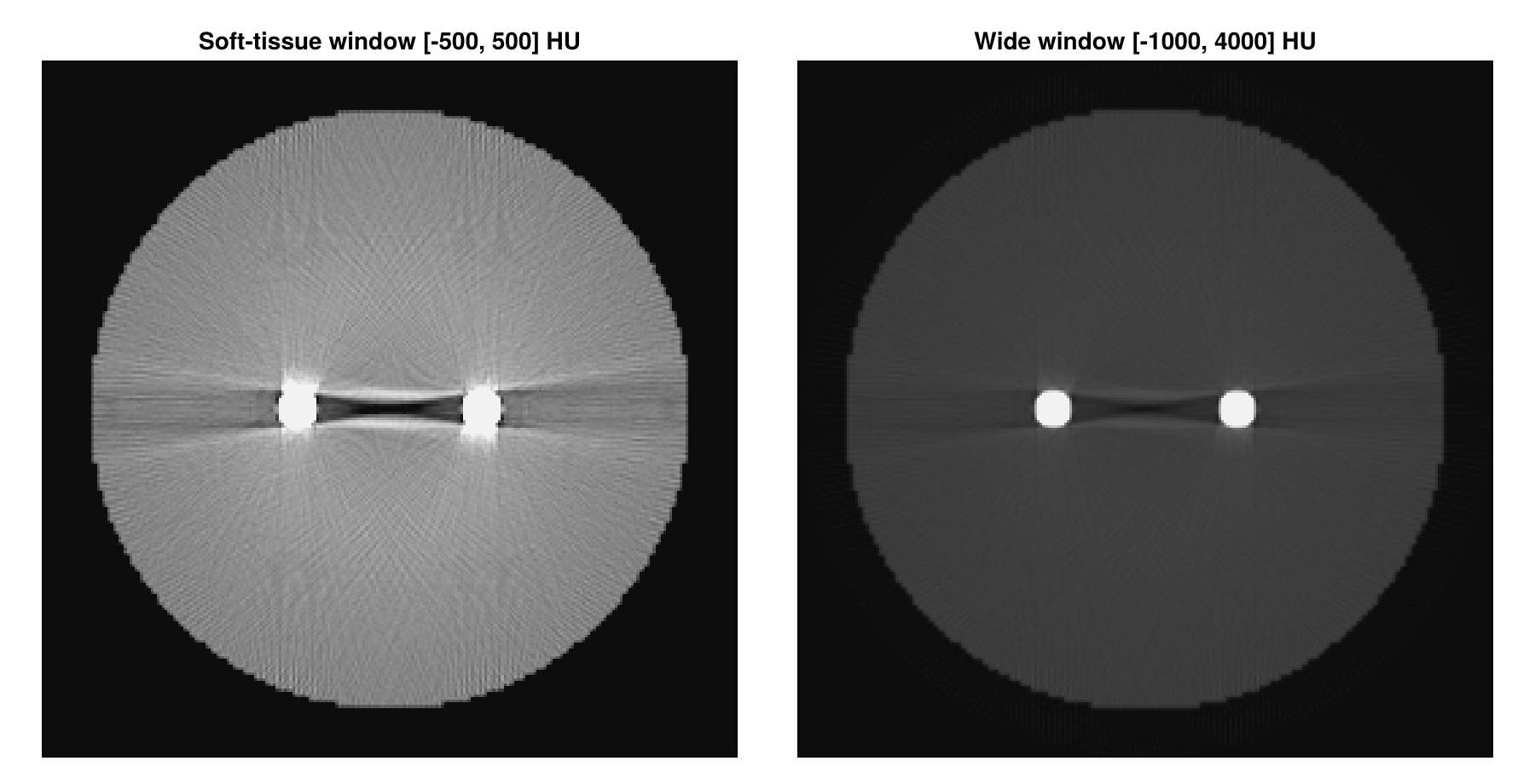
Titanium defined as a user material and two rods scanned in water at 120 kVp: the dark band of beam hardening and the streaks of photon starvation through the standard water BHC and FDK chain, measured against the same scan without the metal.
CatSim vs BasisSimulator
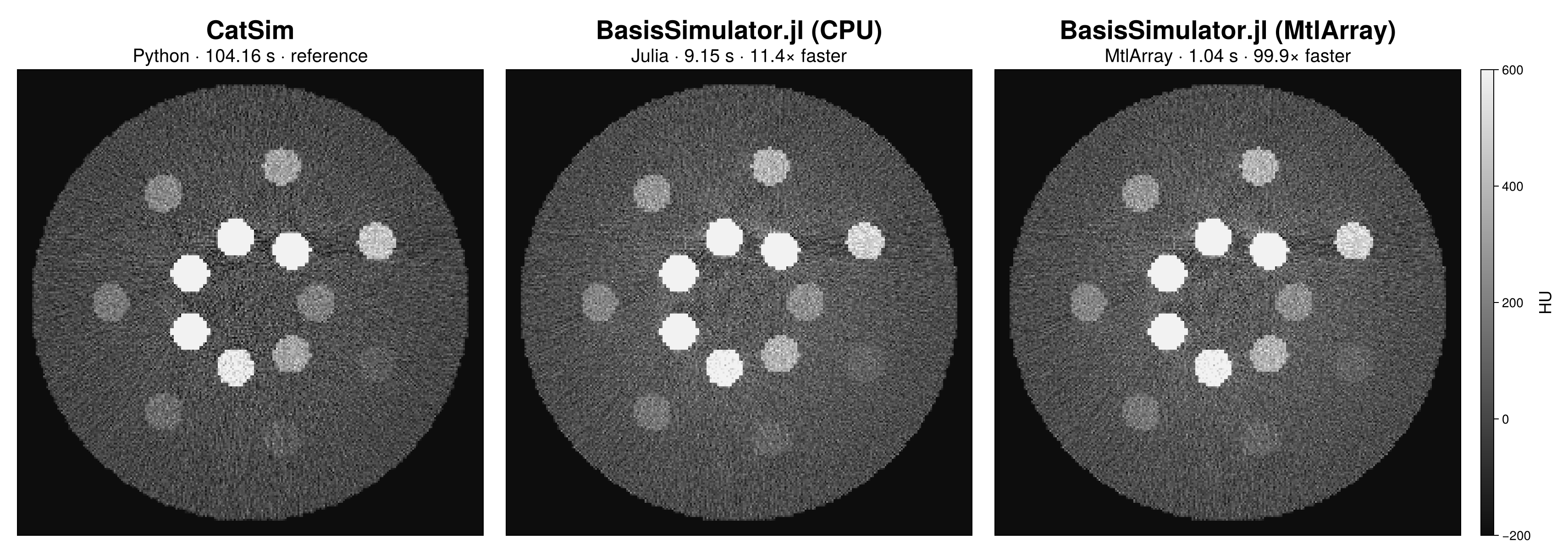
The same Gammex 472 scan through CatSim/XCIST and BasisSimulator on the CPU and the GPU, configured to the same beam and water BHC: images, differences, per-rod HU agreement against theory, and the timing of each pipeline.